Home

millimeter Spectacle Svig skin histology h&e prototype Græsse Tablet

Histology Laboratory Manual

Histology of Skin Stained with H&E Stock Photo - Image of skin,  hematoxylin: 42769166
Histology of Skin Stained with H&E Stock Photo - Image of skin, hematoxylin: 42769166

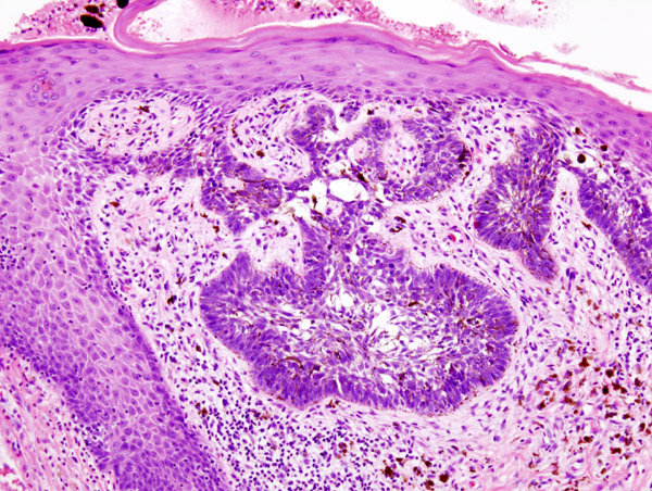
20.Skin (3) Systemic lupus erythematosus (SLE)|Pathology Core Pictures

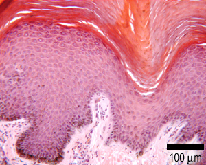
Human Heavily Pigmented Skin sec. 7 m H&E Stain Microscope Slide:  Amazon.com: Industrial & Scientific
Human Heavily Pigmented Skin sec. 7 m H&E Stain Microscope Slide: Amazon.com: Industrial & Scientific

H E Stain: Most Up-to-Date Encyclopedia, News & Reviews

Hematoxylin & eosin (H&E) staining of micropig skin. The histology is... |  Download Scientific Diagram
Hematoxylin & eosin (H&E) staining of micropig skin. The histology is... | Download Scientific Diagram
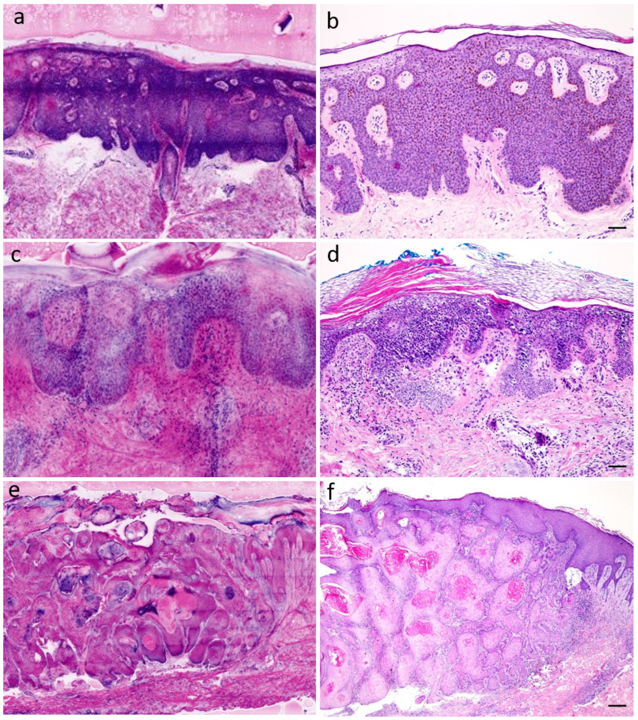
HistoQuarterly: SKIN | Histology Blog

Dermatopathology. Special stains and tests | DermNet

Diagnostics | Free Full-Text | A Feasibility Study for Immediate  Histological Assessment of Various Skin Biopsies Using Ex Vivo Confocal  Laser Scanning Microscopy
Diagnostics | Free Full-Text | A Feasibility Study for Immediate Histological Assessment of Various Skin Biopsies Using Ex Vivo Confocal Laser Scanning Microscopy

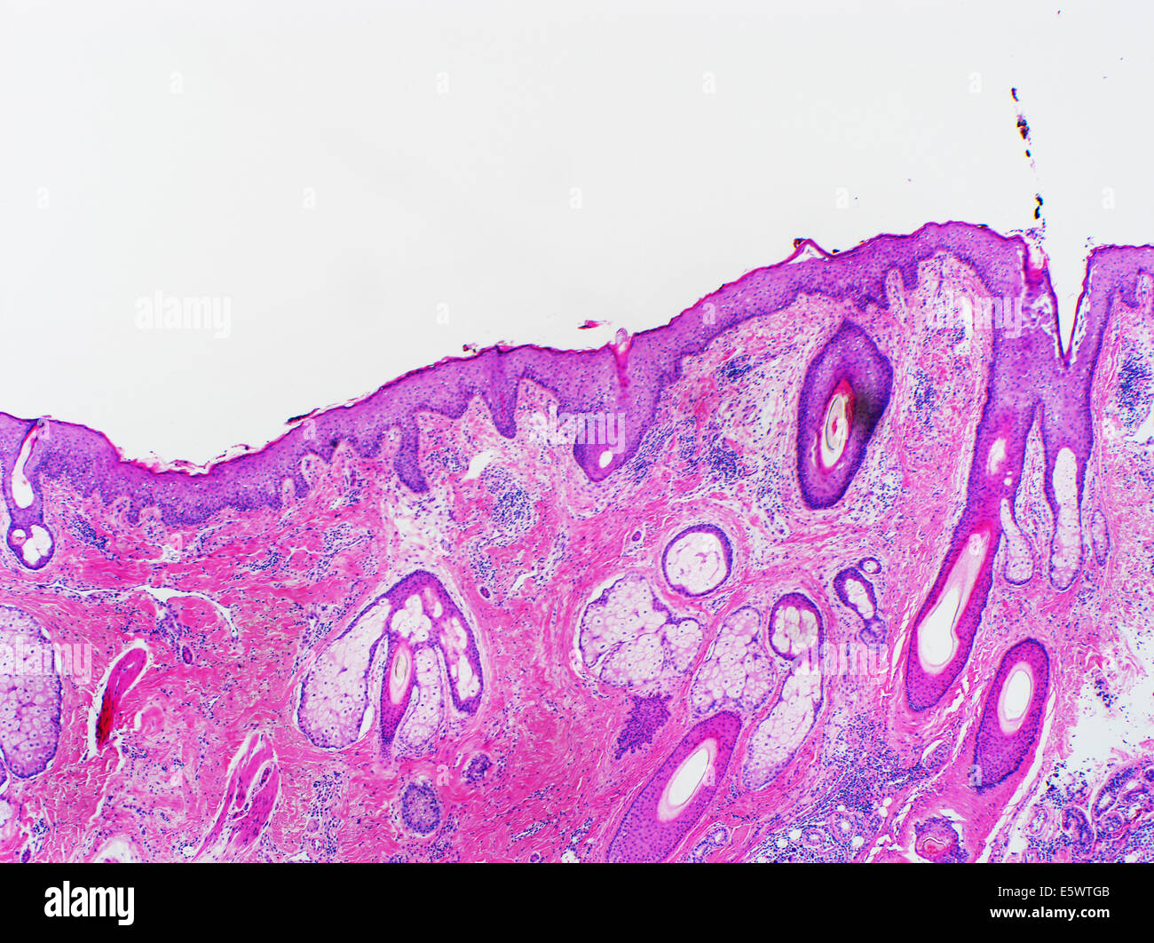
Hematoxylin and eosin stain hi-res stock photography and images - Alamy

Skin: The Histology Guide

Histomorphology and Vascular Lesions in Dorsal Rat Skin Used as Injection  Sites for a Subcutaneous Toxicity Study - Monique Y. Wells, Hélène Voute,  Valérie Bellingard, Cécile Fisch, Virginie Boulifard, Catherine George,  Philippe
Histomorphology and Vascular Lesions in Dorsal Rat Skin Used as Injection Sites for a Subcutaneous Toxicity Study - Monique Y. Wells, Hélène Voute, Valérie Bellingard, Cécile Fisch, Virginie Boulifard, Catherine George, Philippe

Tip Chapter: Histology and Physiology of the Skin | SpringerLink

HistoQuarterly: SKIN | Histology Blog

A novel method for tissue segmentation in high-resolution H&E-stained  histopathological whole-slide images - ScienceDirect
A novel method for tissue segmentation in high-resolution H&E-stained histopathological whole-slide images - ScienceDirect

Figure 5 from Oral nanotherapeutics: Redox nanoparticles attenuate  ultraviolet B radiation-induced skin inflammatory disorders in Kud:Hr-  hairless mice. | Semantic Scholar
Figure 5 from Oral nanotherapeutics: Redox nanoparticles attenuate ultraviolet B radiation-induced skin inflammatory disorders in Kud:Hr- hairless mice. | Semantic Scholar

Histomorphology and Vascular Lesions in Dorsal Rat Skin Used as Injection  Sites for a Subcutaneous Toxicity Study - Monique Y. Wells, Hélène Voute,  Valérie Bellingard, Cécile Fisch, Virginie Boulifard, Catherine George,  Philippe
Histomorphology and Vascular Lesions in Dorsal Rat Skin Used as Injection Sites for a Subcutaneous Toxicity Study - Monique Y. Wells, Hélène Voute, Valérie Bellingard, Cécile Fisch, Virginie Boulifard, Catherine George, Philippe

Histological and immunohistochemical examination of mouse skin treated with  and without imiquimod cream: (A) hemoxylin and eosin (H&E) staining; (B)  the epidermal thickness measured by H&E staining; (C) CD3+ T cell staining;
Histological and immunohistochemical examination of mouse skin treated with and without imiquimod cream: (A) hemoxylin and eosin (H&E) staining; (B) the epidermal thickness measured by H&E staining; (C) CD3+ T cell staining;

Figure 4: Tissue section from submitted tissue, non-haired skin. H&E stain,  20x. | eClinpath
Figure 4: Tissue section from submitted tissue, non-haired skin. H&E stain, 20x. | eClinpath

Skin H&E - Saffron Scientific Histology Services

Normal skin histology. Histology shows epidermis (E), dermis (D) and... |  Download Scientific Diagram
Normal skin histology. Histology shows epidermis (E), dermis (D) and... | Download Scientific Diagram
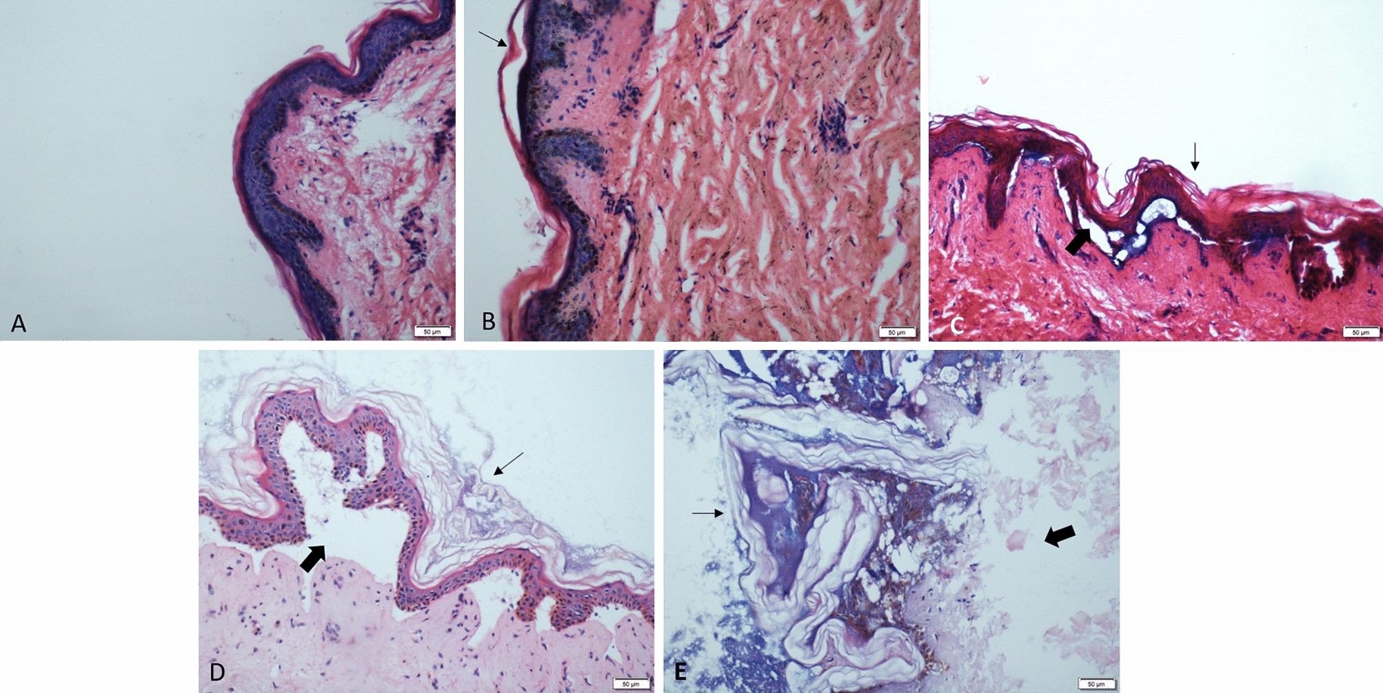
Histological changes in human skin 32 days after death and the potential  forensic significance | Scientific Reports
Histological changes in human skin 32 days after death and the potential forensic significance | Scientific Reports

H&E stain - Wikipedia